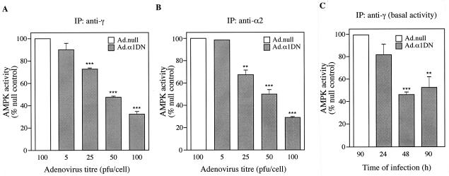
FIG. 5.
Expression of α1DN inhibits AMPK activity. (A and B) Hepatocytes were infected with varying titers of Ad.α1DN or Ad.null. Forty-four hours after infection, AICA riboside (500 μM) was added to the culture medium, and the hepatocytes were incubated for a further 1 h. AMPK complexes were isolated from cell lysates by immunoprecipitation (IP) with either an anti-γ antibody (A) or an anti-α2 antibody (B). Activity present in the immune complexes was measured by phosphorylation of the SAMS peptide. For panel C, hepatocytes were infected with either Ad.α1DN (10 PFU/cell) or Ad.null (10 PFU/cell) in the absence of AICA riboside. At various times postinfection, AMPK activity present in immune complexes isolated using an anti-γ antibody was determined as described above. In each case, results are plotted as a percentage of the activity present in hepatocytes infected with Ad.null and are the mean ± the SEM of four independent experiments. ∗∗∗ denotes a significant difference from the Ad.null value (P < 0.0005), and ∗∗ indicates that P was <0.005.

