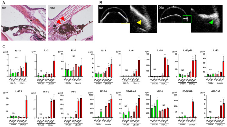
Figure 2.
Age-dependent increase of cytokine levels in AqH in DBA2J mice. (A) The angle structure and iris tissue were normal in DBA2J at 8 weeks (* Schlemm’s canal); however, peripheral synechiae (PAS), iris nodules and iris atrophy developed at 50 weeks (red arrows). Scale bars: 50 μm. (B) In vivo anterior segment optical coherence tomography showed the absence of PAS at 8 weeks (white arrowheads), whereas PAS developed at the age of 50 weeks (green arrowhead). (C) The AqH levels of IL-2, IL-5, IL-6, IL-10, IL-12p70, IL-13, IL-17A, IFN-γ, TNF-α, MCP-1, PDGF-BB and GM-CSF were significantly elevated at 50 and 100 weeks in DBA2J, compared to 8 weeks in DBA2J. * p < 0.05, ** p < 0.001.

