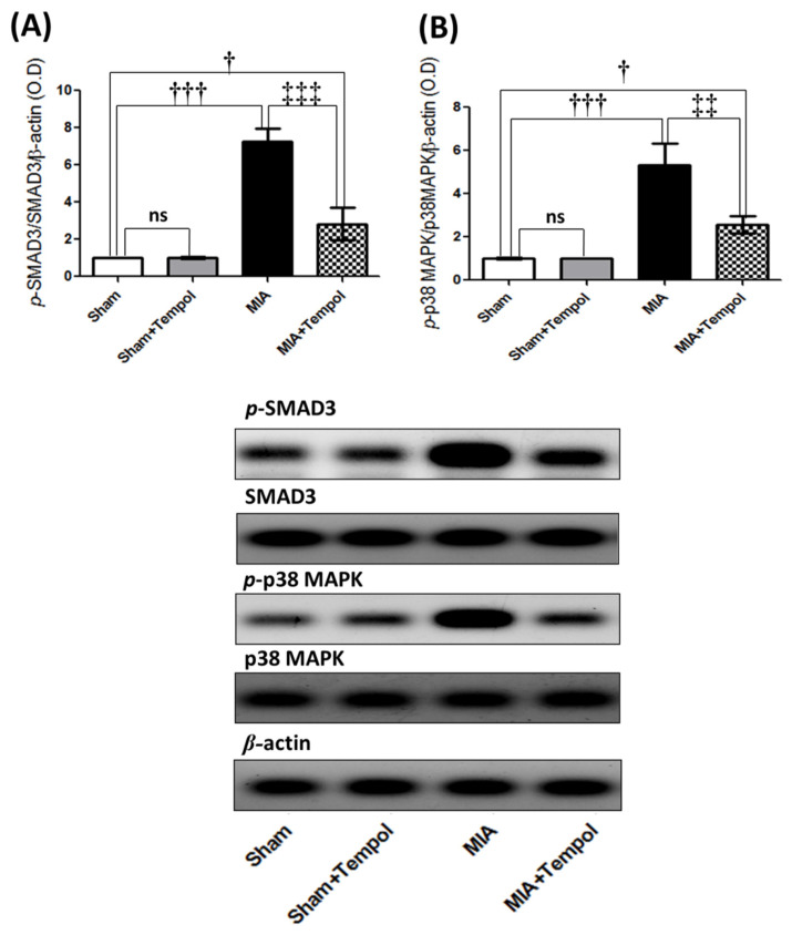
Figure 5.
Effect of Tempol intracellular signaling of p-SMAD3 and p-p38MAPK on osteoarthiritic rats. Rats were subjected to a single intra-articular injection of 3 mg MIA/50 μL saline in their right knees, and then tempol was administered starting from the 7th day of the experiment in a dose of (100 mg/kg/day) by oral gavage for 21 consecutive days. Values of (A) The protein expression of the phosphorylated small mother against decapentaplegic 3 homologs (p-SMAD3), and (B) The protein expression of phosphorylated p38 mitogen-activated protein kinase (p-p38MAPK). The cropped blots of p-SMAD3 and p-p38MAPK were presented relative to that of β-actin, and the uncropped images are available in the Supplementary File. Statistical analysis was carried out using one-way ANOVA followed by Tukey’s multiple comparison test. Results are expressed as mean ± SD (n = 3). † p < 0.05 and ††† p < 0.001 vs. the sham group, and ‡‡ p < 0.01 and ‡‡‡ p < 0.001 vs. MIA group.

