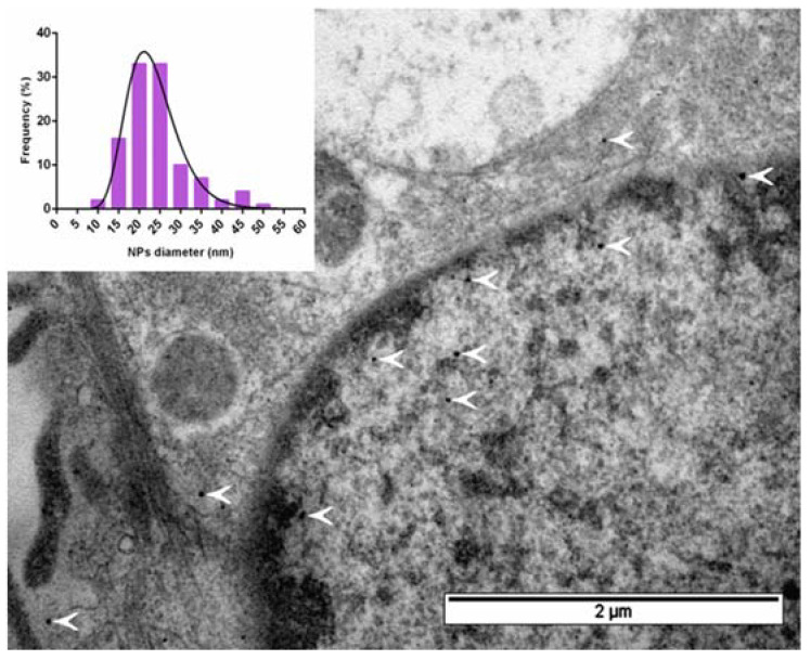
Figure 5.
Bright-field TEM image of an ultrathin section of hAD-MSCs-Mx cells after 24 h of incubation with 2 mM FAS; white arrows indicate iron oxide deposits inside encapsulin nanocompartments. The inset illustrates the size distribution of iron oxide cores inside the encapsulin shell. Scale bar 2 µm.

