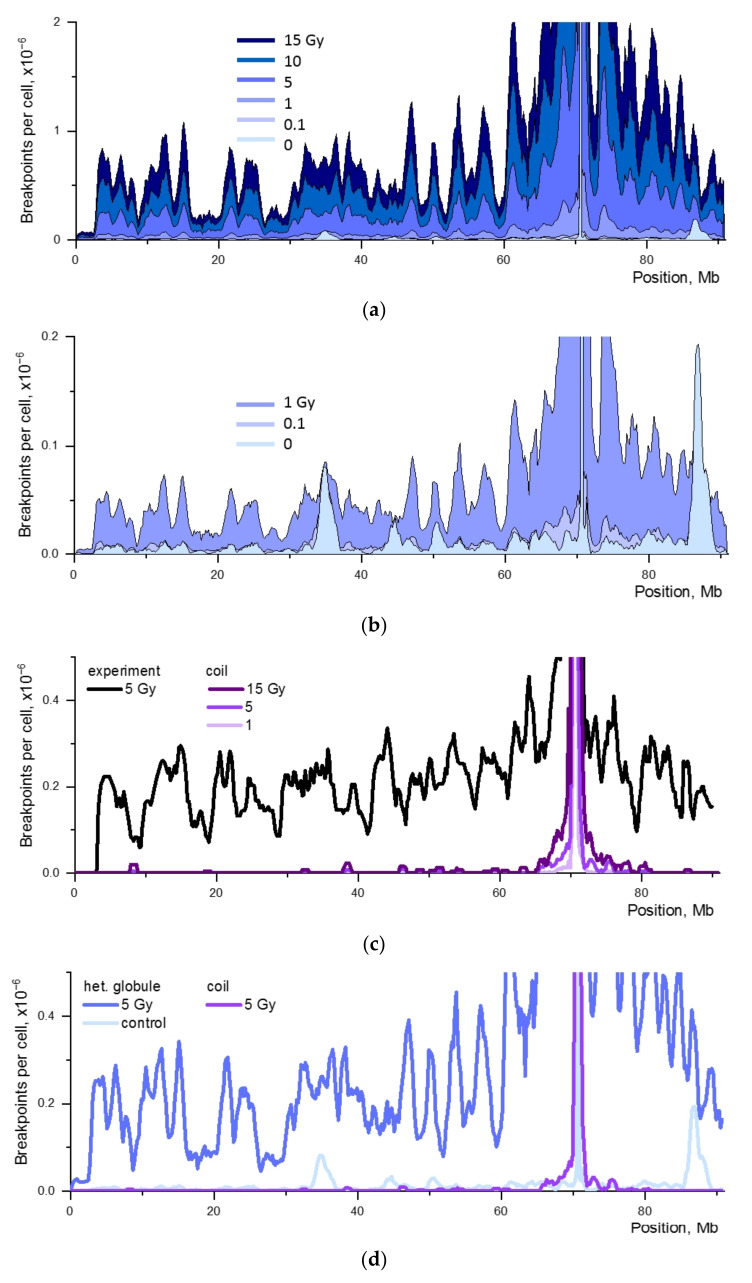
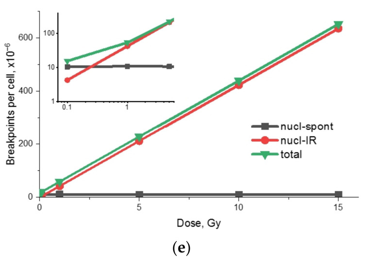
Figure 4.
The impact of targeted and nontargeted DSBs on the formation of cis-translocation breakpoints. The calculations were made for chromosome 18 and the contact-first mechanism. Pc-e = 0.0029. (a,b) The chromosome model is a heteropolymer globule. Dose dependence of the breakpoint distribution. (a) Doses of 0–15 Gy. (b) Low doses (0–1 Gy) shown on an enlarged scale. (c) The breakpoint distribution for a polymer coil model, doses of 1–15 Gy. The experimental translocation breakpoint distribution for chromosome 18, D = 5 Gy [21]. (d) Breakpoint distribution for a heteropolymer globule and a polymer coil models of chromosome 18. D = 5 Gy and D = 0 (simulation for a heteropolymer globule model). (e) Dose response for the frequency of breakpoints of different origin. Abbreviations: nucl-spont, aberrations formed by the nuclease-induced and the spontaneous DSB; nucl-IR, aberrations formed by the nuclease-induced and the IR-induced DSB. The inset shows the low-dose range.


