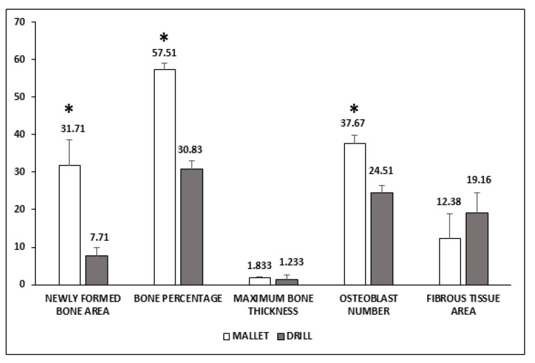
Figure 3.
Histological evaluation of peri-implant bone tissues. Areas of newly formed bone and fibrous tissues are expressed as mm2 and have been evaluated using an imaging computer software. Maximum bone thickness is expressed as mm and was evaluated, as the osteoblast number, by optical microscopy. Data represent the mean ± SEM of the evaluations carried out on bone samples obtained at T14. Student’s t-test: Newly Formed Bone Area * p = 0.031; Bone Percentage * p = 0.001; Osteoblast number * p = 0.009.

