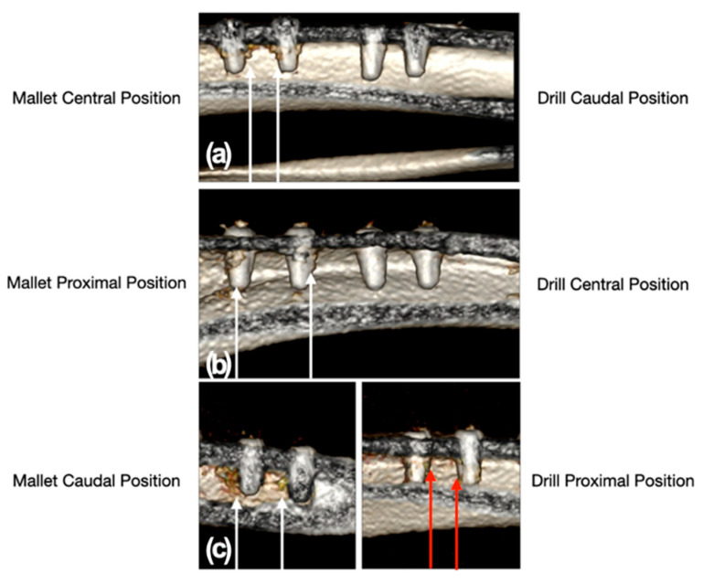
Figure 5.
CT scan images of the position of the implant sites relative to the tibial bone. Panel (a): mallet in central and drill in caudal position; panel (b): mallet in proximal and drill in central position; panel (c): mallet in caudal and drill in proximal position. The white arrows indicate the two sites, with the relative implants, prepared with the mallet technique. In these sites, a moderate trabecular densification organized at the cortico-cancellous junction adjacent to all implants could be noted. The sites prepared with drill showed no trabecular bone densification adjacent to the implants, except in one site where a slight, nonorganized trabecular densification with the presence of radiodense millimeter spots was present (red arrows).

