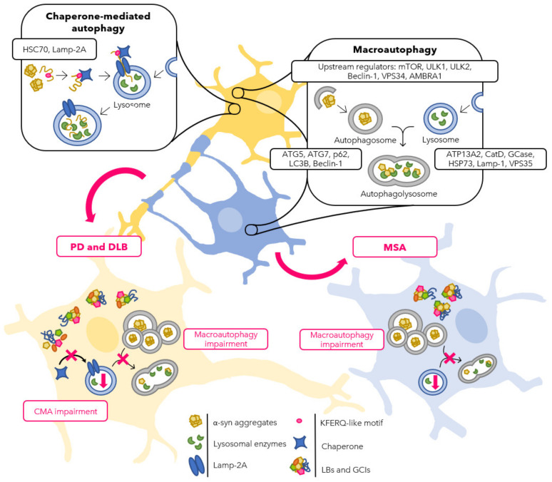
Figure 2.
Schematic representation of the autophagic flux in neurons (yellow) and oligodendrocytes (blue) in normal and PD, DLB, and MSA conditions. Physiologically, both CMA and macroautophagy pathways are involved in protein degradation in the cytoplasm of neurons, whereas macroautophagy seems to be the more effective mechanism in oligodendrocytes. It has been reported that, in the context of ASPs, an impairment of the autophagic machinery is involved. PD and DLB neurons (bottom left) present with LBs in the cytoplasm accumulating α-syn together with other proteins that can be degraded by neither macroautophagy nor CMA, such as LRRK2, DJ-1, and UCHL1. Moreover, other autophagy-related proteins, such as AMBRA1, p62, and LC3B, accumulate in LBs. Eventually, macroautophagy impairment results in an accumulation of autophagosomes and reduction in lysosomal activity. MSA oligodendrocytes (bottom right) show defects as well in the macroautophagic pathway with accumulation of GCIs containing α-syn, AMBRA1, p62, LC3B, and HSC70, together with an increased number of autophagosomes and diminished degradation in the lysosomes.

