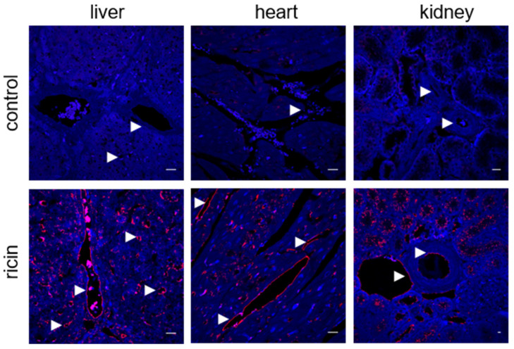
Figure 3.
Ricin binding to swine tissues. Immunofluorescence (red) of the liver, heart and kidney sections before (control) or 30 min after incubation with ricin (5 µg/mL). The arrows indicate blood vessels and ricin binding. Nuclei stained by DAPI appear in blue. Scale bar: 20 µm (representative sections of n = 3 swine/group are shown).

