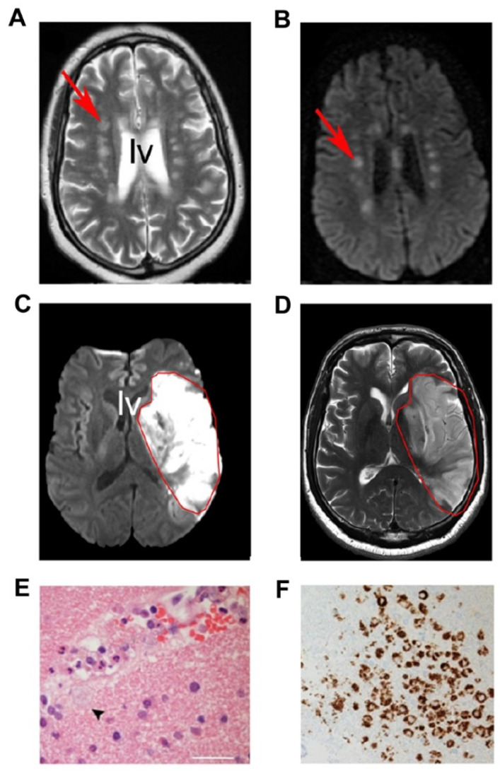
Figure 4.
CNS pathology in COVID-19 victims. (A,B) MRI showing small foci of injuries (arrows) near the lateral ventricle (LV) and SVZ. (C,D) Large lesion (outlined in red) near the lateral ventricles. (E) A small blood vessel surrounded by immune cells that invaded the brain. Note macrophage extending into brain (small arrowhead). (F) CD68 immunohistochemistry showing macrophages around small vessels. (Adapted from Paterson et al., 2020), with permission.

