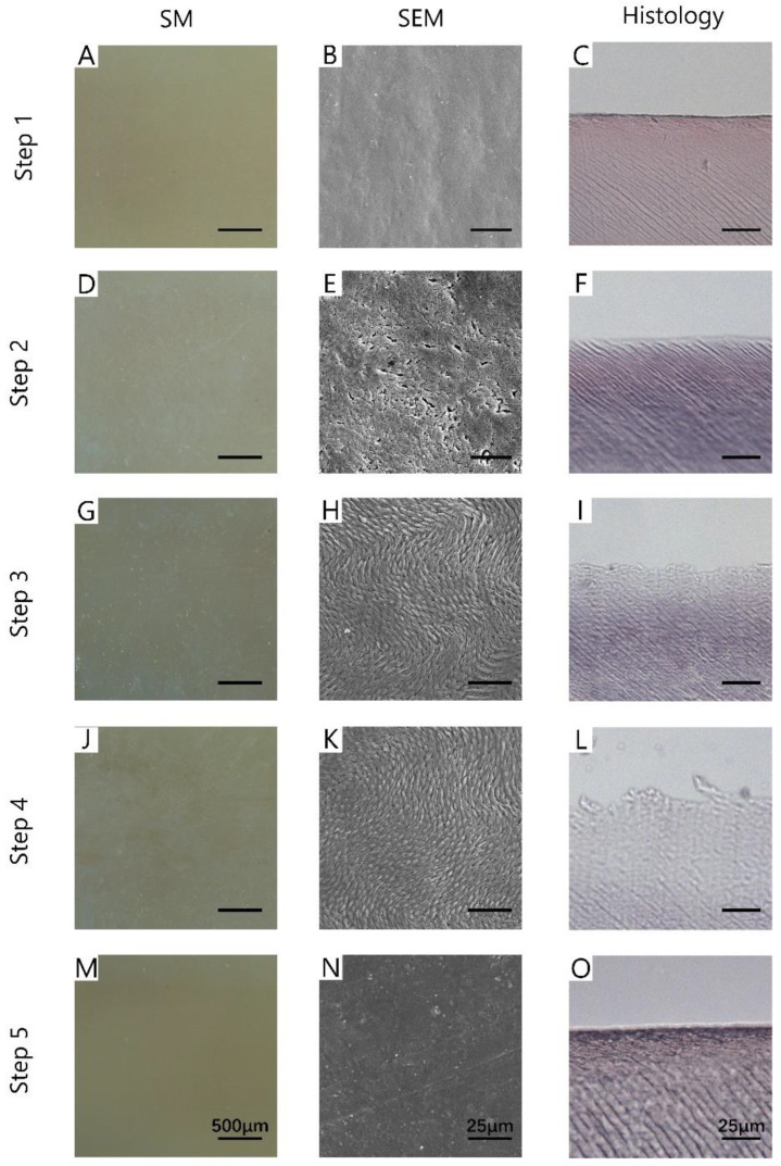
Figure 4.
The images of SM (A,D,G,J,M), SEM (B,E,H,K,N), and histology (C,F,I,L,O) of each step. Step 1: the surface of the incisor was clean; Step 2: 37% phosphate etching agent was evenly applied to the incisor for 15 min, after which the surface was clean and dried; Step 3: Icon-Etch was smeared evenly on the surface of incisor for 30 s, after which the surface was clean and dried; Step 4: Icon-Dry was smeared evenly on the surface of incisor and kept for 1 min, after which the surface was dried; Step 5: Icon-Infiltrant was smeared evenly on the surface of maxillary incisor for 1 min and light-cured for 40 s. Scale bar of SM is 500 μm; scale bar of SEM is 25 μm; scale bar of histology is 25 μm.

