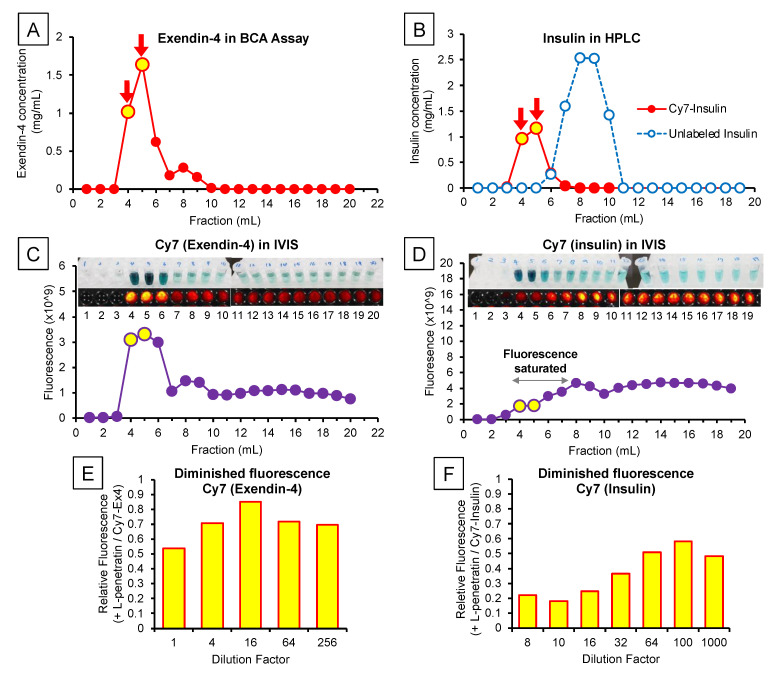
Figure 3.
Characterization of the peptides labeled with a fluorescent probe (Cy7). Panels (A) and (B), respectively, show the concentrations of Exendin-4 (BCA assay) and insulin (HPLC) in the eluted fractions after labeling with Cy7. Panels (C,D) show the fluorescence intensity in the eluted fractions measured by the in vivo imaging system and the reference photos of these samples. The fluorescence intensity of fractions 4–7 of Cy7-labeled insulin exceeded the upper limit of detection, in contrast with the visible fluorescence shown in the photos (panel (D)). Panels (E,F) indicate the weakened fluorescence of Cy7-labeled Exendin-4 and insulin mixed with L-penetratin. L-penetratin reduced the fluorescence of Cy7-labeled Exendin-4 and insulin to 54% and 22% of that without L-penetratin, respectively. Dilution allowed partial fluorescence recovery to 70% and 48%.

