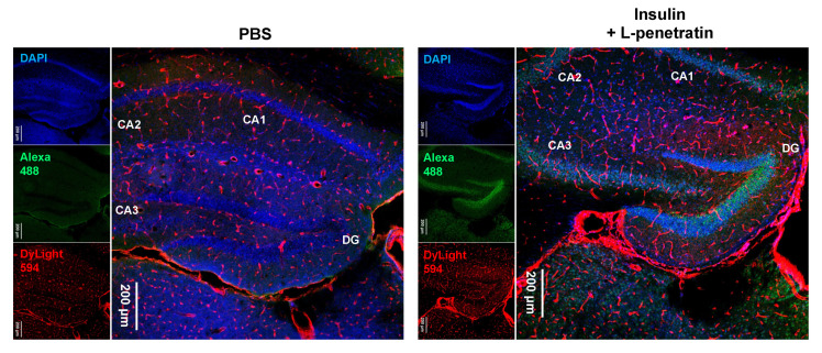
Figure 7.
Immunohistological staining of the hippocampus at −1.91 mm from bregma after intranasal administration of PBS or insulin with L-penetratin to mice. Insulin was detected with an anti-insulin primary antibody and Alexa 488-conjugated secondary antibody (green). Blood vessels were stained with DyLight 594 conjugated tomato lectin (red). The images show a typical result obtained in one experiment. Separate experiments under the same conditions yielded similar results (n = 3 or 4).

