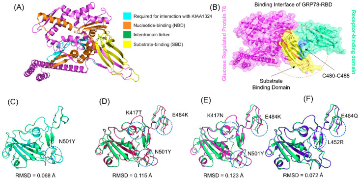
Figure 1.
Structural modeling and analysis of GRP78 and spike RBD of the wild-type and docking interaction analysis B.1.1.7, P.1, B.1.351, and B.1.617 variants. (A) shows the structure of GRP78 and its domain organization; (B) shows the binding interface of GRP78 and spike RBD; (C) shows the superimposed structure of wild-type and B.1.1.7 variant RBD; (D) shows the superimposed structure of wild-type and P.1 variant RBD; (E) shows the superimposed structure of wild-type and B.1.1.7 variant RBD; (F) shows the superimposed structure of wild-type and B.1.617 variant RBD.

