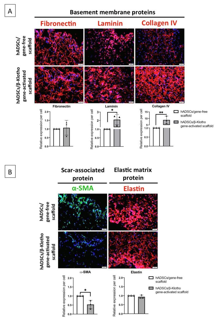
Figure 4.
Deposition of pro-wound healing matrix proteins by the ADSCs in the gene-activated scaffold on day 14. (A) The ADSCs in the gene-activated scaffold demonstrated significant regeneration of the basement membrane compared to the ADSCs in the gene-free scaffold. The ADSCs predominantly deposited a relatively mature network of collagen IV proteins, followed by laminin and fibronectin. (B) The ADSCs in the gene-activated scaffold also expressed 2-fold lower of the α-SMA protein compared to the ADSCs in the gene-free scaffold. In conjunction with basement membrane regeneration, the ADSCs in the gene-activated scaffold deposited qualitatively better elastin matrix. All the images were captured through 20× objective using an IX73 Olympus microscope. ** and * indicates p < 0.01 and p < 0.05 respectively. Scale bar 50μm. Data represents mean ± standard deviation (n = 3). α-SMA and fibronectin was double-immunostained.

