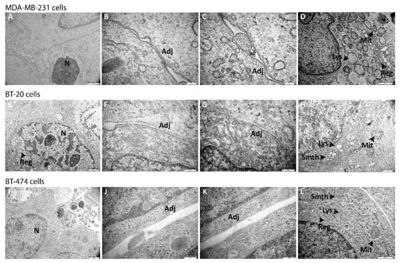
Figure 4.
Ultrastructure of MDA-MB-231, BT-20, and BT-474 spheroids by transmission electron microscopy (TEM). Images of (A–D) MDA-MB-231, (E–H) BT-20, and (I–L) BT-474 spheroids formed under optimized conditions were acquired after 7 days of culture with TEM microscope Hitachi H-7000. Scale bars are shown on all images. N = nucleus; Adj = adjoined connections; Mit = mitochondria; Lys = lysosome; Reg = rough endoplasmic reticulum; Smth = smooth endoplasmic reticulum.

