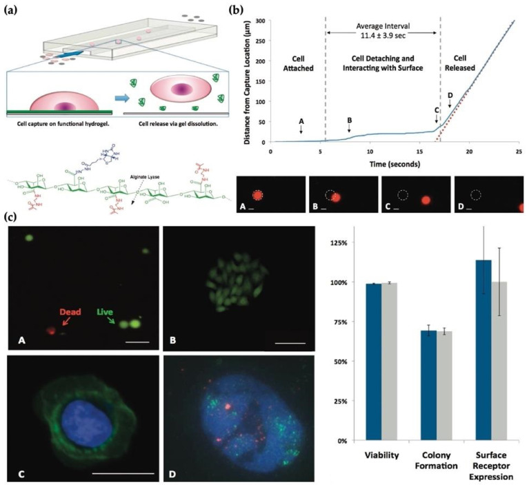
Figure 1.
(a) Sacrificial hydrogel coatings microfluidic devices; (b) The gentle nature of the release process as the cell starts; (c) released cells for (A) viability using a fluorescent LIVE (green)/DEAD (red) assay and (B) colony formation, (C) immunostaining of cell surface receptors, (D) FISH (fluorescence in situ hybridization) analysis in a released HER2 (green probe) amplified breast cancer cell; the control probe (red) [78].

