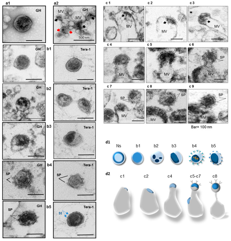
Figure 3.
Morphology of HML.2 GH and HML.2 Tera-1 isolated from sucrose gradient fractions with a buoyant density 1.16 g/cm3. Demonstration of viral elements capture by presumed MVs. Particles were examined by negative staining (a) and by ultrathin section (b,c) TEM. Cartoons of capsids and envelopes of the virus-like particles (d1) and MVs with captured viral cargo (d2). (a1) Negative staining. Bald particles with amorphous capsids were detected in HML.2 GH preparation. (a2) HML.2 GH preparation that was not filtered through the 0.45 µm membrane but purified by sucrose Dgc. Obtained pellets were diluted and analyzed. Aggregate composed of extracellular vesicles with attached virus-like particles (red arrows) and dense virus-like content inside vesicles are shown (black arrows); (b) comparative analyses of HML.2 GH and HML.2 Tera-1 free virions; (b1) bald virion with amorphous capsid; (b2) bald virion with fragmented capsids; (b3) bald virions with condensed ovoid eccentric capsids; (b4) virions with SP and partially condensed capsids; (b5) virion with SP (left) and virion with partially condensed capsid with presumed TM stumps (St, blue arrows) (right); (c) capture of viral elements (black arrows) at different steps of assembly by presumed MVs; (c1,c2) supposed Gag complexes under the cell membrane (early budding stage); (c3–c5) assembly of viral capsid (mid budding stage); (c5) SP at the surface of particle in formation; (c6) assembled particle, constriction of the neck before completion of the budding (late budding stage); (c7) three virions at different stages of assembly trapped inside overlapped MVs. Particle (upper right) with constriction neck, condensed capsid and SP (late budding stage); (c8) assembled virion with SP attached to MV; (c9) supposed free virion with SP and amorphous capsid. Bars represent 100 nm. MV: Microvesicle. SP: spike proteins (or surface projections) at the surface; (d1) graphical interpretation of obtained results. Morphology of HML.2 free particles from teratocarcinoma cells (upper panel). Immature particles with toroidal capsids are not shown (Ns). Particle with amorphous capsid is shown in light blue and condensed capsids are given in dark blue; (d2) variants of HML.2 viral cargo in MVs. Virions with SP (shown as “Y”) and condensed capsids were not frequent. Virions with vRNA (shown as a “spring”) are not frequent.

