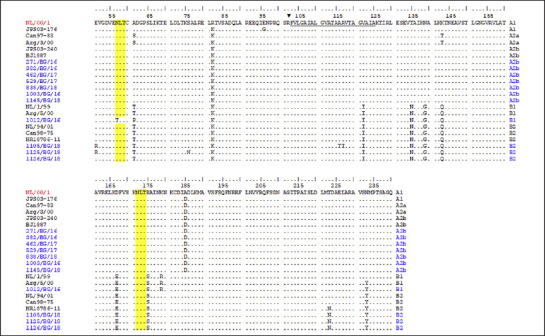
Fig. 3.
Deduced amino acid alignment of partial F protein of hMPV strains. The alignment is shown relative to the sequence of reference NL/00/1 strain (GenBank accession number: AF371337). The Bulgarian hMPV strains are indicated in blue. Identical residues are identified as dots. The fusion peptide is underlined. The protease cleavage site is indicated with a black triangle. Yellow shading highlights the predicted N-glycosylation sites. hMPV, human metapneumovirus.

