Figure 7.
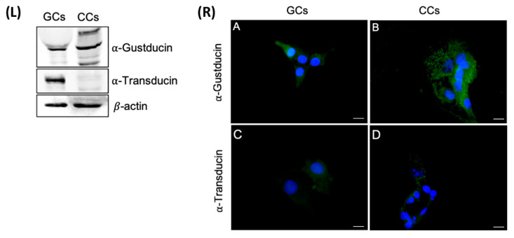
Left panel (L) The upper panel shows the detection of α-gustducin by Western blot with an anti-α-gustducin antibody, in CCs and GCs protein extracts. The central panel shows the detection of α-transducin by Western blot with an anti-α-transducin antibody in GCs and CCs protein extracts. β-actin was used as loading control. Right panel (R) Immunofluorescence localization of (A,B) α-gustducin, (C,D) α-transducin, in (A,C) granulosa and (B,D) cumulus cells. α-gustducin and α-transducin are stained in green. Nuclei were counterstained with DAPI (blue). Negative controls are shown in Figure S1. Scale bar = 15 µm.

