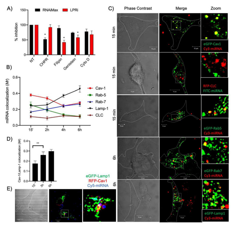
Figure 3.
Intracellular trafficking of miRNA-133a. (A) U87MG LentiRILES/133 T cells were pre-incubated with specific inhibitors of the caveolae (Fillipin and Genistein), clathrin (Chlorpromazine, CHPR) and macropinocytosis (cytochalasin D, Cyto D) internalization pathways before miRNA-133a transfection with LPRi or RNAiMax. Then, 48 h later, the luciferase activity in cells was quantified (n = 3). (B) U87MG cells expressing eGFP-Cav1, RFP-CLC, eGFP-Rab5, eGFP-Rab7 or eGFPLamp1 were transfected for 1 h (pulse) with LPRi.Cy3-miRNA-133a then washed and further incubated for 15 min, 2, 4 or 6 h, fixed in 3% PFA and analyzed by confocal microscopy analysis (n = 4). (C) Representative images from eGFP-Cav1, RFP-CLC and eGFP-Rab5 cells collected at a 15-min time point and from eGFP-Rab7 or eGFP-Lamp1 cells collected at a 6 h time points. (D) Quantification of co-localization events between RFP-Cav1 and eGFP-Lamp1 detected at 15 min, 3 and 6 h postincubation (n = 3). (E) Co-localization of LPRi.Cy5-miRNA-133a with RFP-Cav1 and eGFP-Lamp1 after 3 h incubation (n = 3). For all panels, values are mean ± SEM; * p < 0.05, ** p < 0.01; Reproduced with permission from Elsevier [79].

