
| Although prefrontal areas have been widely studied and implicated in various brain functions and disorders, there is a surprising lack of commonly accepted nomenclature and delineation of its subdivisions. Rodent stereotaxic atlases, on which experimentalists rely the most, are regularly updated as no consensus is found [15]. In the absence of clear landmarks to define the mPFC, a lot is left to individual appreciation which can lead to apparent incoherencies between studies and overall misinterpretation. Until a unified nomenclature is accepted in the field, it is necessary that authors report precise stereotaxic coordinates and explicitly define the brain region(s) they study. | |
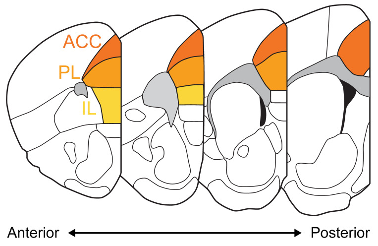
For simplicity here, we will use the following
nomenclature for the 3 major subdivisions of the mPFC:
| |
| Although the human PFC evolved to be relatively bigger and more complex than the rodent PFC, notably with more clearly defined subregions, homologies in embryological development, layer organization, cell-type distribution and connectivity patterns advocate for potentially shared functions. For an anatomical definition and a comparison between human and rodent PFC, see Carlén, 2017 [16]. For a very detailed description of the cytoarchitecture of the mouse PFC, see Van de Werd et al., 2010 [17], and for a comparison between mouse reference atlases, see Le Merre et al., 2021 [15]. | |
|
Box figure. Coronal sections of the mouse brain along the antero-posterior axis with the 3 major subdivions of the mPFC highlighted (Anterior cingulate cortex ACC, Prelimbic cortex PL, Infralimbic cortex IL) based on the Allen Brain Atlas. |
Official websites use .gov
A
.gov website belongs to an official
government organization in the United States.
Secure .gov websites use HTTPS
A lock (
) or https:// means you've safely
connected to the .gov website. Share sensitive
information only on official, secure websites.