Figure 7.
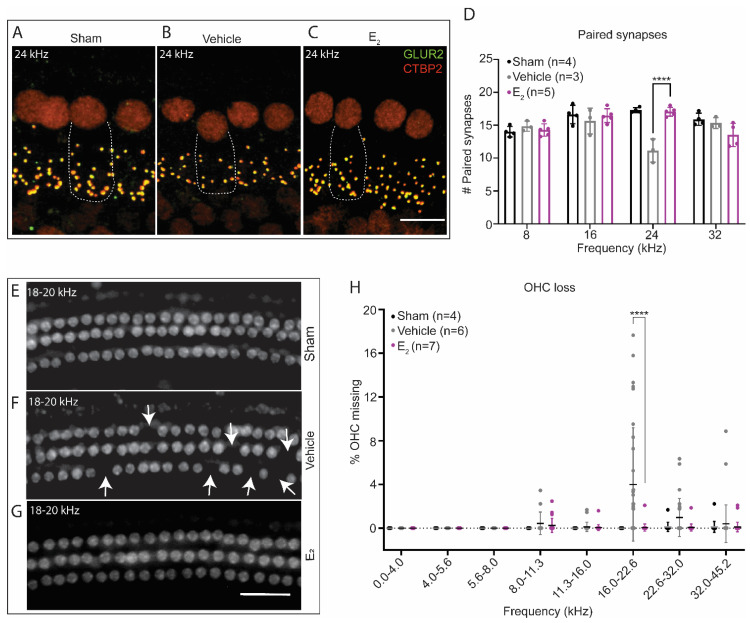
E2-replacement in gonadectomized female mice ameliorates cochlear synaptopathy and OHC loss following a TTS-inducing noise exposure. (A–C) Representative images of paired IHC-ANF synapses at 24 kHz 1-week post-exposure in (A) sham-exposed mice, (B) vehicle-treated mice, and (C) E2-treated mice. Paired synapses were identified via the co-localization of CTBP2 (red) and GLUR2 (green). Sham animals were gonadectomized and vehicle-treated but not noise-exposed. (D) E2-replacement (150 μg/kg) ameliorates cochlear synaptopathy at 24 kHz 1-week following a TTS-inducing noise (94 dB SPL, 8–16 kHz, 2-h) (p < 0.0001). Compared to a group of sham exposed mice, E2-treated mice display no loss of synapses at 24 kHz, while vehicle-treated mice display, on average, a loss off 5.9 synapses at this frequency. (E–G) Representative cytocochleograms 1-week post-exposure at the approximate cochlear location of 18–20 kHz in (E) sham-exposed mice, (F) vehicle-treated mice, and (G) E2-treated mice. White arrows indicate missing OHCs. (H) Vehicle-treated mice display increased susceptibility to OHC loss 1-week following a TTS-inducing noise exposure. Compared to E2-treated female mice, vehicle-treated female mice displayed significantly increased OHC loss restricted to the frequencies between 16.0 kHz and 22.6 kHz (p < 0.0001). Neither group of animals displayed significant OHC loss below 16 kHz or above 22.6 kHz. Paired synapse counts and OHC loss were compared using a 2-way ANOVA followed by Sidak’s multiple comparison test. Dotted lines in (A–C) represent the outline of a single inner hair cell. n = number of ears, plots: mean ± SD; scale bar in (C): 10 µm; scale bar in (G): 50 µm; **** p < 0.0001.

