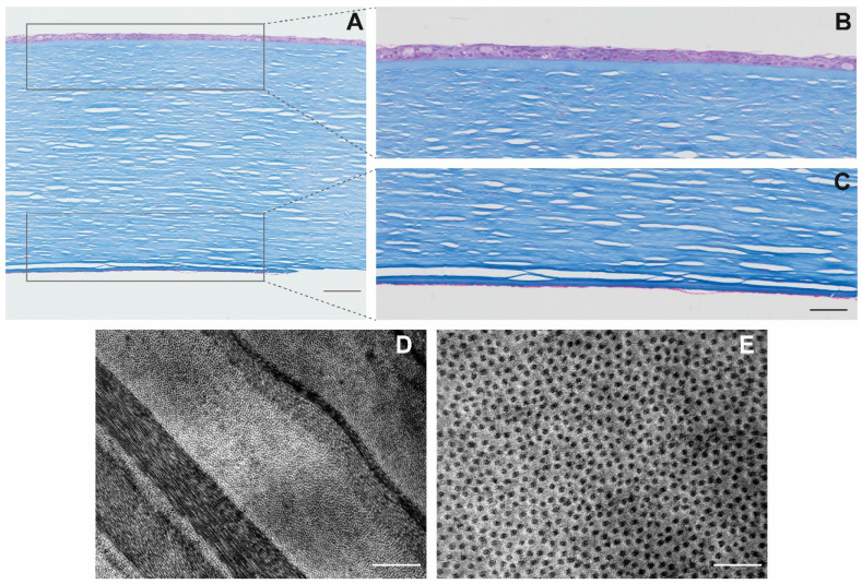
Figure 2.
Transversal cuts of TK-cryopreserved corneas stained with Masson’s trichrome. All corneal layers are visible (A) (scale: 100 μm). Details of the epithelium (B) and the endothelium (C) (scale: 50 μm). TEM image of the banded pattern of collagen fibers in P1-cryopreserved corneas (D) (scale: 1 μm), with the details visible in a transversal cut of the collagen fibers (E) (scale: 200 nm).

