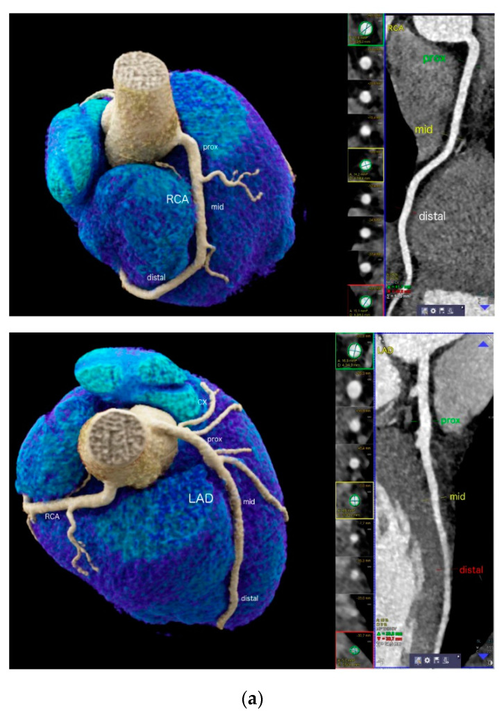
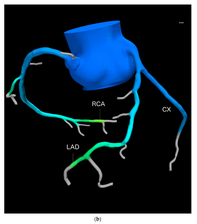
Figure 1.
(a) 22-year-old professional male endurance athlete (swimmer) with regular high training volumes and a new onset of high-grade arrhythmia. Enlarged proximal but smaller mid and distal RCA (upper) and LAD (lower panel) segments (“athlete’s artery”). Quantitative CTA (right) included sizing of RCA and LAD dimensions at 3 sites (proximal, mid and distal) using curved multiplanar reformations (cMPR). Both vessel area (mm2) and 2 diameters (mm) were taken. Left panels show volume rendering technique (VRT). CX = circumflex artery. (b) 3D Quantification of vessel volumes (mm3) for the RCA, LAD and CX by using a computational fluid modelling (CFD) software (Heartflow, Inc. Redwood, CA, USA) in another patient.


