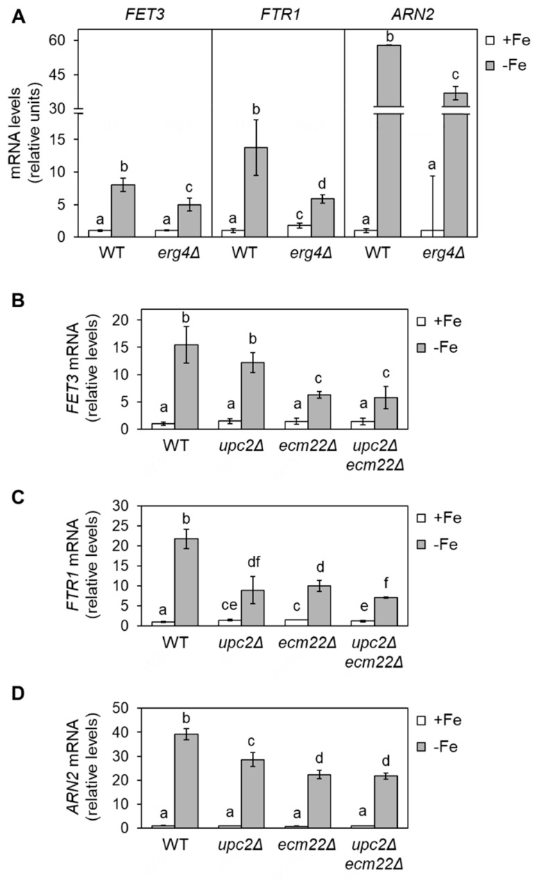
Figure 2.
Defects in ergosterol biosynthesis limit the up-regulation of iron regulon genes during iron deficiency. (A) Wild-type (WT, BY4741) and erg4Δ cells were cultivated in SC at 30 °C in SC medium without (+Fe) or with 100 µM BPS (−Fe) for 6 h. (B–D) Wild-type (WT, W303), upc2Δ, ecm22Δ, and upc2Δecm22Δ cells were grown for 15 h to exponential phase in SC medium (+Fe) and then 100 µM BPS was added, and cells were cultivated for 9 h (−Fe). Total RNA was extracted, and mRNA levels of FET3, FTR1, and ARN2 were determined by RT-qPCR as indicated in Material and Methods. Data were normalized to PGK1 (A) or ACT1 (B–D) mRNA levels. Data display the average and standard deviation (SD) of three biologically independent assays relative to wild-type cells in +Fe conditions. Different letters above bars indicate statistically significant differences (p-value < 0.05).

