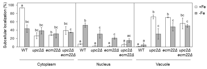
Figure 7.
Upc2 and Ecm22 are necessary for the correct localization of Aft1 protein. Quantitative analysis of Aft1 subcellular localization patterns in wild-type (WT, W303), upc2Δ, ecm22Δ, and upc2Δecm22Δ cells containing JK1346 (pRS426-GFP-AFT1) plasmid. Cells were cultivated as indicated in Figure 6 and visualized under Nomarski (DIC) and GFP fluorescence optics. More than 100 cells were counted for at least three different experiments. GFP-Aft1 distribution patterns were considered as cytoplasmic, nuclear or vacuolar. Average and SD were represented. Statistical analysis has been performed independently for cytoplasm, nucleus, and vacuole, and different letters above bars of each panel indicate significant differences (p-value < 0.01).

