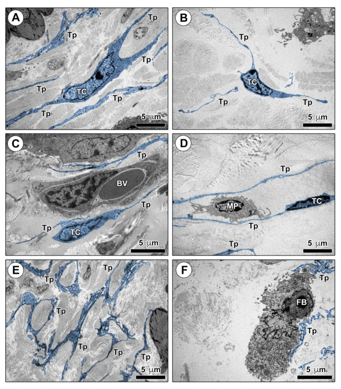
Figure 4.
Ultrastructural identification of telocytes (TCs) in control mouse skin. (A–F) Representative transmission electron microscopy photomicrographs of skin ultrathin sections from control mice stained with UranyLess and bismuth subnitrate solutions. TCs and telopodes have been digitally colored in blue. TCs are ultrastructurally identifiable as stromal cells characterized by (i) a spindle-shaped, oval, or piriform cell body with a relatively large euchromatic nucleus surrounded by a scarce cytoplasm, and (ii) the presence of telopodes, long cytoplasmic processes with a narrow emergence from the cell body and a moniliform appearance due to the alternation of thin segments (podomers) and expanded portions (podoms). TCs are widely distributed in control mouse dermis, where their long telopodes surround collagen bundles (A,B) and blood microvessels (C), and establish intercellular contacts with macrophages (D). Telopodes often form a labyrinth-like network distributed among dermal collagen bundles (E) and contact fibroblasts (F). Scale bar: 5 μm (A–F). BV, blood vessel; FB, fibroblast; MP, macrophage; TC, telocyte; Tp, telopode.

