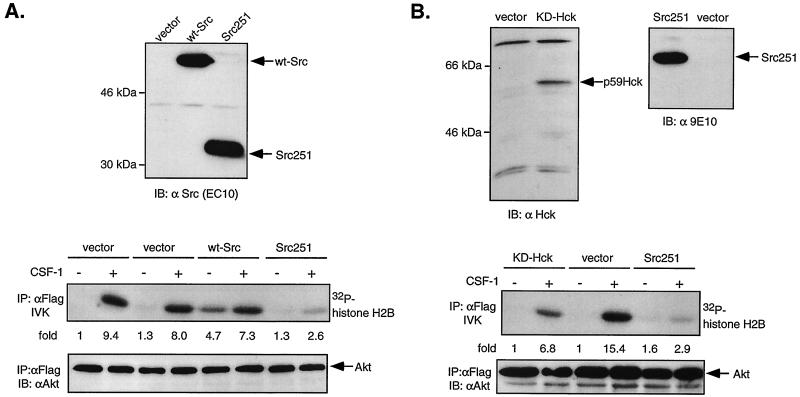
FIG. 8.
Dominant-negative SFKs inhibit CSF-1-stimulated Akt activity in ΔKI-CSF-1R cells. (A) ΔKI-CSF-1R cells were transiently transfected with either 21 μg of pEF-BOSΔR1 vector and 1 μg of Flag-Akt, 20 μg of pcDNA vector and 2 μg of Flag-Akt, 20 μg of WT Src and 2 μg of Flag-Akt, or 20 μg of Src251 and 2 μg of Flag-Akt. Cells were starved and stimulated with 10 nM CSF-1 for 2 min. (Top) Total cell lysates were immunoblotted (IB) with an anti-Src antibody. (Middle) Lysates were immunoprecipitated (IP) with anti-Flag antibodies and analyzed for histone H2B kinase activity. (Bottom) The amount of immunoprecipitated Flag-Akt was detected with anti-Akt antibodies. (B) ΔKI-CSF-1R cells were transiently transfected with 2 μg of Flag-Akt and 20 μg of either vector (pEF-BOSΔR1), KD-Hck, or Src251. (Top) Total cell lysates were analyzed for Hck expression with an anti-Hck antibody and for Src251 expression with anti-Myc antibody. (Middle and bottom) Cells were processed as described for panel A.

