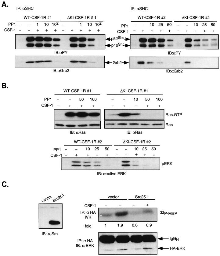
FIG. 9.
Role of SFKs in activating the Ras/ERK pathway in ΔKI-CSF-1R cells. (A) Shc tyrosine phosphorylation and association with Grb2. WT clones 1 and 2 and ΔKI clones 1 and 2 were pretreated with PP1 at the concentration (micromolar) as indicated and stimulated with 10 nM CSF-1 for 2 min. Lysates were immunoprecipitated (IP) with anti-Shc antibodies, separated by SDS-PAGE, and transferred to a membrane. The top part was immunoblotted (IB) with anti-PY antibodies, and the bottom part was probed with anti-Grb2 antibodies. (B) Ras and ERK activation. (Top) WT clone 1 and ΔKI clone 1 cells were pretreated with PP1 at the concentration (micromolar) indicated and stimulated with 10 nM CSF-1 for 2 min. Ras activation was assayed by binding 500 μg of lysates to GST-RBD. Total Ras was determined in 50 μg of total cell lysates. (Bottom) WT clone 2 and ΔKI clone 2 cells were treated as indicated, and total cell lysates were analyzed with anti-active ERK antibodies. (C) Effect of dominant-negative Src expression. ΔKI-CSF-1R cells were transiently transfected with 5 μg HA-ERK2 and 20 μg of either vector or Src251. Lysates were analyzed for MBP kinase activity (IVK) and blotted for ERK levels in anti-HA immunoprecipitates. This experiment has been repeated twice with similar results.

