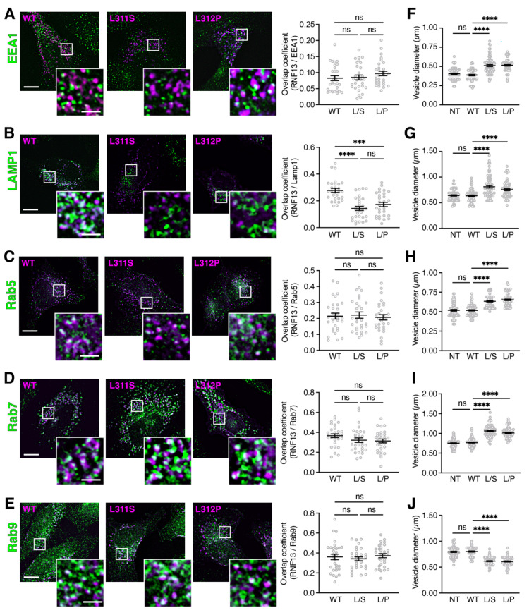
Figure 1.
RNF13 L311S and L312P variants alter the size of endolysosomal vesicles. (A–E) Plasmid constructs encoding HA-tagged WT, L311S, or L312P RNF13 were transiently expressed in HeLa cells. Cells were fixed, permeabilized, and labeled with specific primary antibody against HA (RNF13) and against the endogenous subcellular markers. Representative images from three independent experiments (N = 3) show RNF13-HA (in purple) and (A) early endosome marker EEA1, (B) lysosome marker Lamp1, (C) early endosome marker Rab5, (D) late endosome marker Rab7, and (E) recycling endosome marker Rab9 (in green). Scale bars indicate 10 µm for whole cell image and 2 µm for the boxed area shown at higher magnification. The jittered individual value plots (A–E) represent Manders overlap coefficient of RNF13 over (A) EEA1, (B) Lamp1, (C) Rab5, (D) Rab7, or (E) Rab9. For each condition analyzed: N = 3, n = 30 cells. Values are expressed as mean ± SEM. Not significant (ns), *** p = 0.0004 or **** p < 0.0001 using one-way ANOVA with the Kruskal–Wallis test. The jitter strip plots ((F), EEA1; (G), Lamp1; (H), Rab5; (I), Rab7; (J), Rab9) represent the diameter of the five largest vesicles for each cell. For each condition analyzed: N = 3, n = 15 cells, 75 vesicles. Values are expressed as mean ± SEM. Not significant (ns) or **** p < 0.0001 using one-way ANOVA with the Kruskal–Wallis test.

