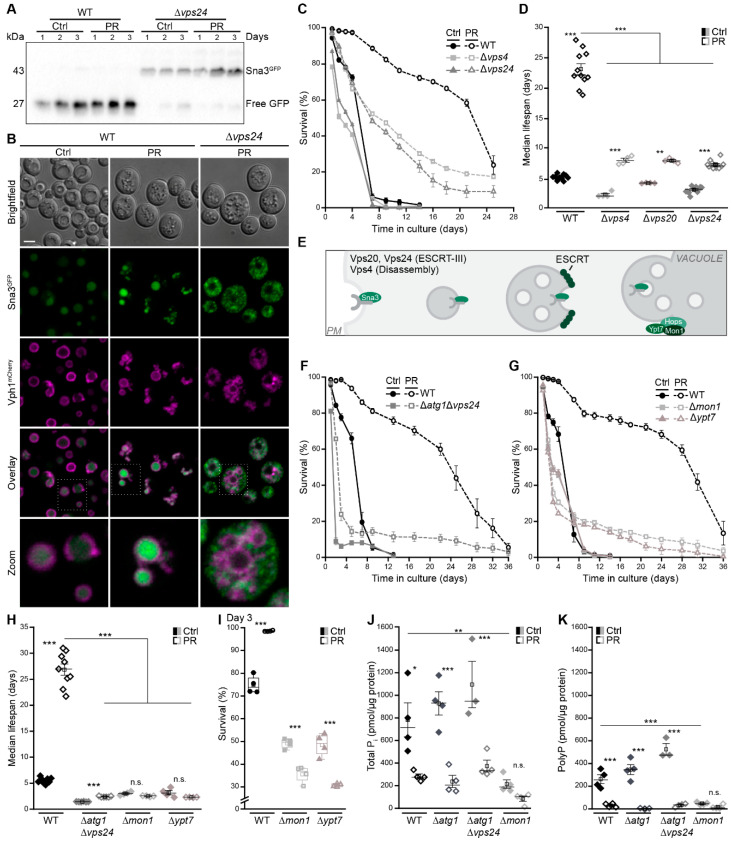
Figure 5.
Autophagy and the multivesicular body pathway work in parallel to sustain survival upon phosphate exhaustion. (A) Immunoblot of WT and ∆vps24 cells harboring endogenously GFP-tagged Sna3 grown in standard (Ctrl) or phosphate-restriction (PR) media. Blots were probed with an antibody against GFP to assess GFP liberation. (B) Confocal microscopy of WT and ∆vps4 cells harboring endogenously GFP-tagged Sna3 and mCherry-tagged Vph1. Images of ∆vps4 cells were post-processed differently to visualize Sna3GFP. Scale bar represents 3 µm. (C) Survival during chronological aging, determined via flow cytometric quantification of PI staining, of WT, ∆vps4 and ∆vps24 cells grown on Ctrl or PR media; n = 4. (D) Median lifespan of WT, ∆vps4, ∆vps20 and ∆vps24 cells. (E) Schematic showing the multivesicular body (MVB) pathway and key players of vacuole fusion. (F–H) Survival during chronological aging of WT and ∆atg1∆vps4 cells (F) and of WT, ∆mon1 and ∆ypt7 cells (G) grown on Ctrl or PR media, as well as corresponding median lifespan (H); n > 4. (I) Survival of WT, ∆mon1 and ∆ypt7 cells on day 3 of the aging shown in (G). (J,K) Total phosphate (J) and polyphosphate (K) levels in WT, ∆atg1, ∆atg1∆vps4 and ∆mon1 cells grown for 24 h on Ctrl or PR media; n = 4, except ∆atg1∆vps4 n = 3. For statistical analysis, Welch-ANOVA with Dunnett T3 post hoc test (D,H) and one-Way ANOVA with Tukey post hoc test (I–K) was used. * p < 0.05, ** p < 0.01 and *** p < 0.001; ns not significant.

