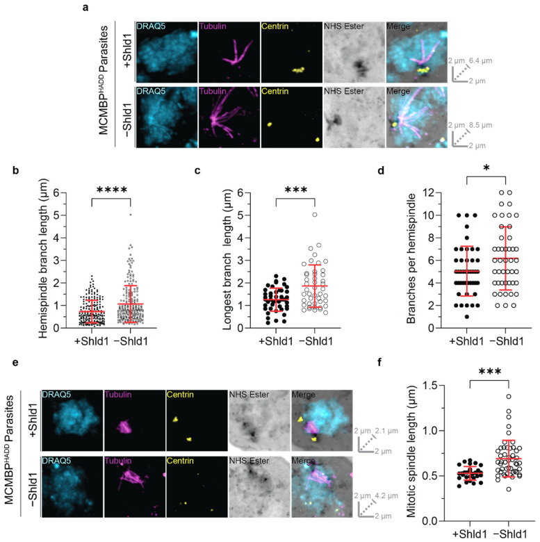
Figure 2.
MCMBP-deficient parasites show defects in both mitotic spindle and hemispindle formation. MCMBPHADD parasites were cultured [+]/[−] Shld1. Parasites were then prepared for U-ExM, stained with a nuclear stain (DRAQ5, in cyan), anti-tubulin (in magenta), anti-centrin (in yellow), and a protein stain (NHS Ester, in grayscale), and visualized using Airyscan microscopy. (a) Hemispindles were imaged, and the length of all hemispindle branches (b), of the longest branch in each individual hemispindle (c), and the total number of branches per hemispindle (d) were all measured. n = 221 hemispindle branches and 45 hemispindles for +Shld1, 214 hemispindle branches and 45 hemispindles for -Shld1 were measured across 3 biological replicates. (e) Mitotic spindles were imaged and their length (f), from one MTOC to another, was measured. n = 28 for +Shld1 and 49 for −Shld1, across 3 biological replicates. All distance measurements presented here have been estimated based on the average expansion factor of gels used in this study. Raw values can be found in Figure S2. (* p < 0.05, *** p <0.001, **** p < 0.0001 by unpaired two-tailed t-test, error bars = SD). All images are maximum intensity projections. Slice-by-slice videos of images are found in Videos S5–S8. Scale bars as labelled in each image, solid bars = XY scale, dashed bar = combined depth of slices used for Z-projection.

