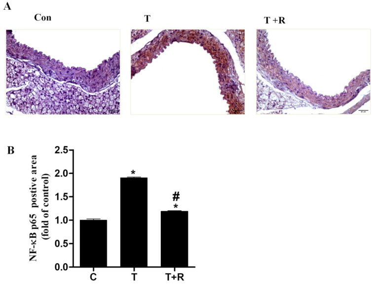
Figure 7.

Representative images showing the immunohistochemical staining for NF-κB p65 in aortic cross-sections (magnification of 40×, scale bar = 50 μm). C57BL/6 mice were fed AIN-93G rodent diets with and without 0.4% resveratrol for one week followed by 25 μg/kg/day of TNF-α injected intraperitoneally for 7 days. After treatment periods, the animals’ aortas were harvested for sectioning. Representative photomicrographs of immunohistochemical staining for NF-κB p65 (A). Quantitative analysis of NF-κB p65 (B). T, TNF-α; T+R, TNF-α + resveratrol. *, p < 0.05 vs. control; #, p < 0.05 vs. TNF-α-alone-treated mice.
