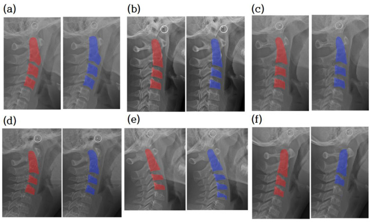
Figure 6.
Cervical vertebrae segmentation mask. Comparison between ground truth of segmentation and prediction. The radiograph overlaid with the ground truth of the segmentation image (red). The radiograph is overlaid with the prediction (blue). (a–d) are examples of accurate segmentation of cervical vertebrae. (e,f) are examples of segmentation of less or more than three vertebrae. Note that cropped images have various sizes.

