Fig. 4.
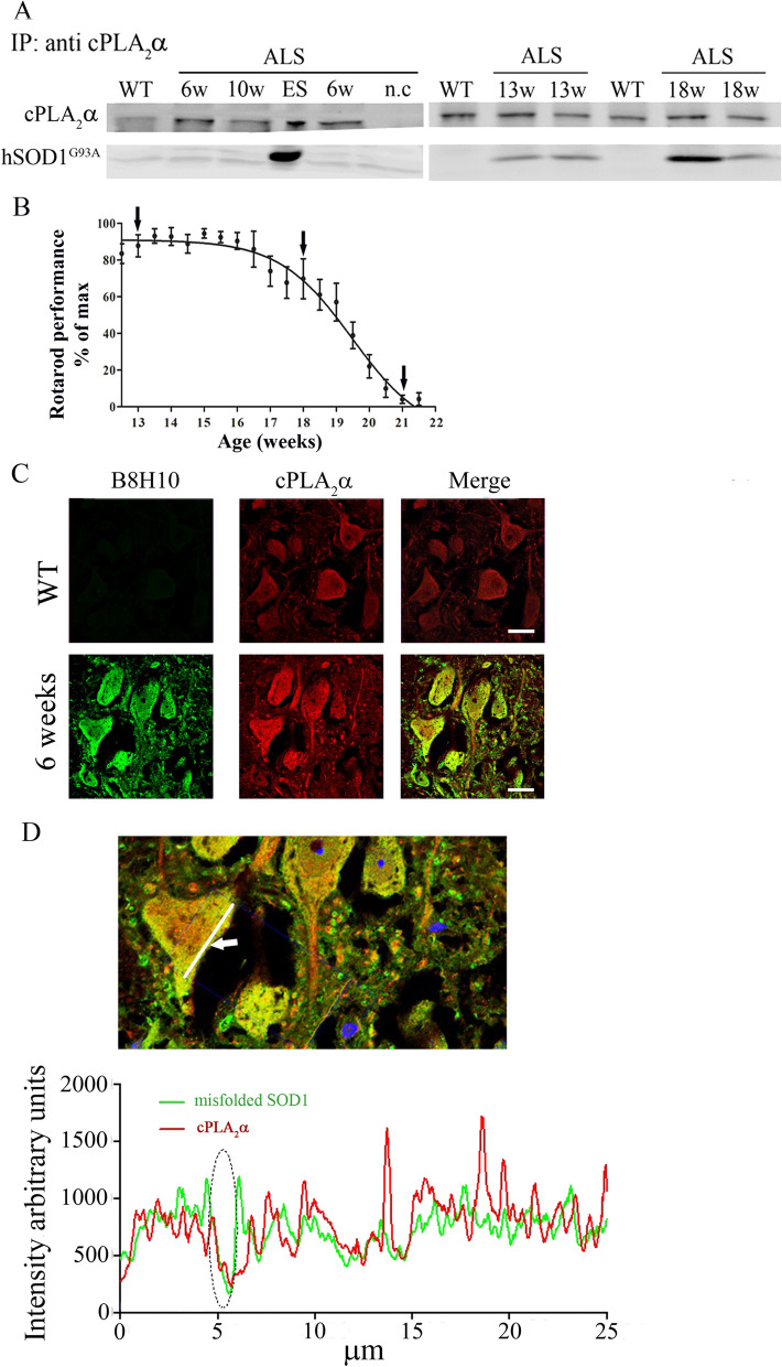
Partial binding between cPLA2α and mutant SOD1G93A in motor neurons of 6 weeks old mutant SOD1G93A mice. A Immunoprecipitation with antibody against cPLA2α and Western Blot analysis for cPLA2α and mutant SOD1 in the spinal cord lysate of control (WT) and mutant SOD1G93A mice during the development of the disease shown by a representative immunoblot. Negative control (n.c). -without antibodies against cPLA2α. B Motor performance on accelerating Rotarod test (n = 10 mice). The arrows show the immunoprecipitation analysis along the development of the disease. C Airyscan detector high resolution confocal microscopy images of double immunofluorescence staining of cPLA2α (red) with misfolded SOD1 (B8H10, green) proteins in the spinal cord sections of 6 weeks old pre-symptomatic SOD1G93A mouse. Scale bars = 20 μm. D The coupling of both proteins (cPLA2α and misfolded SOD1) in the line shown by the arrow was analyzed and presented as intensity along the line. The ellipse shows an example of binding between both proteins

