Fig. 5.
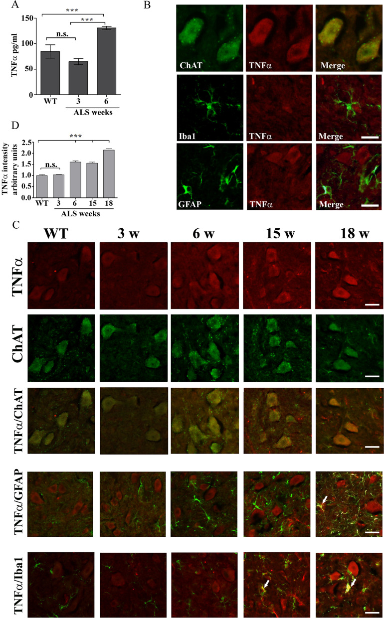
Increased TNFα is restricted to the motor neurons of pre-symptomatic 6 weeks old mutant SOD1G93A mice. A The levels of TNFα in the spinal cord lysate of WT and of 3 and 6 weeks old pre-symptomatic mutant SOD1G93A mice detected by ELISA. Significance—***p < 0.001, n.s. = non-significant. The bar graph is the mean ± SE of 8 mice in each group. B Representative double immunofluorescence staining cell markers (green) of motor neurons (ChAT), microglia (Iba-1) or astrocytes (GFAP) and TNFα (red) in the spinal cord sections of 6 weeks old pre-symptomatic SOD1G93A mice. Scale bars = 20 μm. 3 other mice in each group were analyzed and showed similar results. C A representative time course of double immunofluorescence staining of TNFα (red) and cell markers (green) of motor neurons (ChAT), microglia (Iba-1) or astrocytes (GFAP) proteins in the lumbar spinal cord sections of WT and mutant SOD1G93A mice during the course of the disease (3, 6, 15 and 18 weeks). Scale bars = 20 μm. 3 other mice in each group were analyzed and showed similar results. D. The means ± SEM fluorescence intensity for TNFα is presented in the bar graph as arbitrary units. Four mice for each time point and five fields for each mouse were analyzed. Significance compared to control ***p < 0.001, n.s. non-significant

