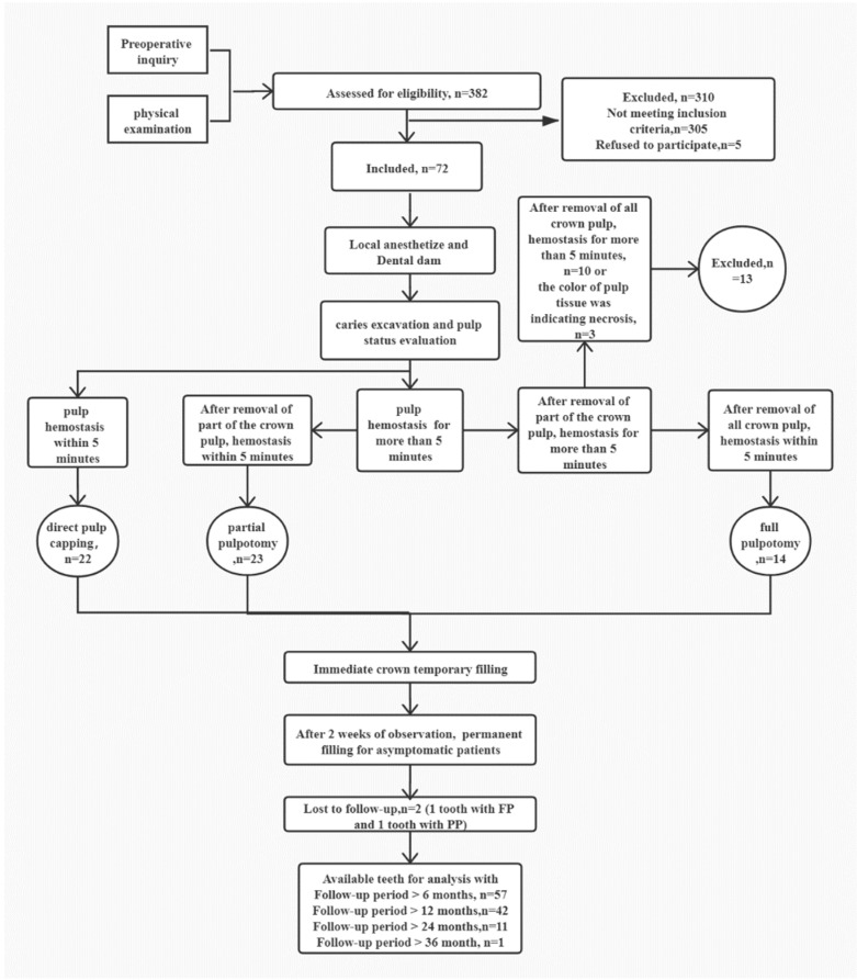
Figure 1.
A schematic diagram of the VPT protocol in this clinical study. The number (n) of patients participating in each stage is given. In general, after anesthetizing and isolating the tooth before caries excavation and pulp exposure, pulp bleeding was assessed, and the pulp status was evaluated under microscope. If pulp hemostasis was achieved by direct contact with a cotton pellet moistened with 1% NaOCl within 5 min, DPC was performed. If the hemostasis was more than 5 min, approximately 2–3 mm or more of affected pulpal tissue underneath the exposure site was removed using a sterile high-speed diamond. After this, if the hemostasis was less than 5 min, PP was performed. Otherwise, the full crown pulp was removed to the level of the root canal orifices. Hereafter, if the hemostasis was less than 5 min, FP was performed. Otherwise, the treated tooth was excluded from the study and further treated with RCT or revascularization according to the root development.

