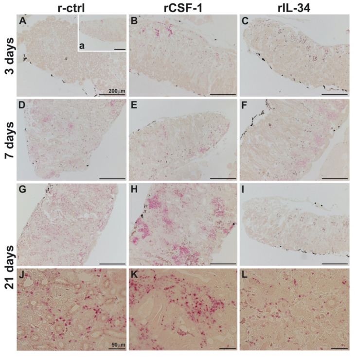
Figure 5.
The kidneys of CSF-1-MΦ-enriched, FV3 infected frogs possess greater infiltration of granulocytes. X. laevis were injected ip with 2.5 μg of rCSF-1 (B,E,H,K) or rIL-34 (C,F,I,L) in APBS or equal volumes of a recombinant vector control (r-ctrl; (A,D,G,J); inset a panel: uninfected) and three days later infected ip with FV3 (5 × 10⁵ PFU). At designated times, animals were sacrificed, and their kidneys were processed for histology and examined by NASDCl-specific esterase (Leder) stain, with granulocytes staining pink. The images are representative of sections from kidneys of four mock-infected and five infected animals per treatment group (n = 4 for uninfected controls and n = 5 for FV3-infected groups). Inset panel in (A), denoted (a) is representative of kidneys from mock-infected animals.

