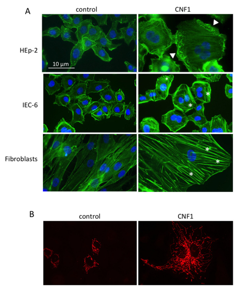
Figure 2.
Example of morphological effects of CNF1 on actin and mitochondria. (A) F-actin and nuclei staining of different cell lines untreated or treated with CNF1. Asterisks: stress fibers; arrow-heads: ruffles. (B) Mitochondrial staining of control and CNF1-treated IEC-6 cells. Note the enrichment of the mitochondrial network in treated cells.

