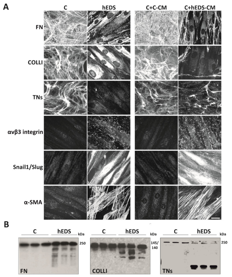
Figure 1.
Proteolytic and differentiation potential of hEDS-CM. (A) On the left: IF analyses of FN-, COLLI, and TNs-ECM organization, αvβ3 integrin and Snail1/Slug transcription factor expression, and α-SMA cytoskeleton assembly in control (C) and patient (hEDS) dermal fibroblasts grown for 10 days (6 days for TNs and COLLI) in complete MEM. The images are representative of 6 different cell strains for each group. On the right: IF analyses of control dermal fibroblasts grown for 10 days (6 days for TNs and COLLI) in the presence of a pool of CM recovered from six 72 h-grown control (C + C-CM) and six hEDS (C + hEDS-CM) cell strains. Images are representative of three independent experiments. Scale bar: 12 μm. (B) WB of 80 µg of proteins recovered from the above-mentioned pooled control and hEDS-CM immunoreacted with anti-human FN Ab, goat anti-human COLLI Ab, and with a mAb recognizing all human isoforms of TNs. WB data represent three technical replicates for each group.

