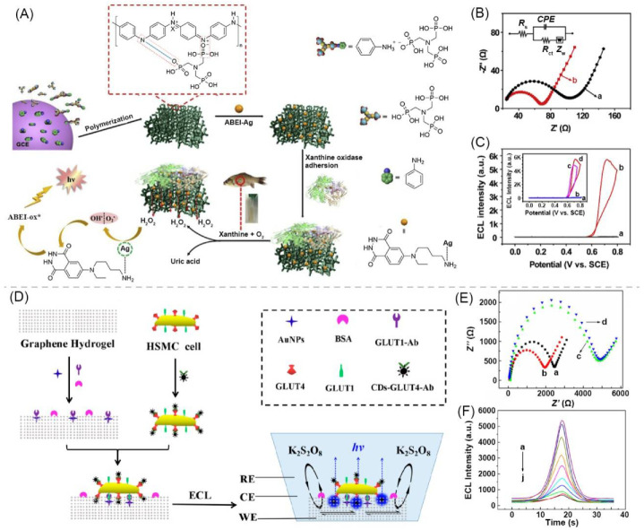
Figure 3.
(A) Schematic of preparation process of PAni polymeric hydrogel-based ABEI–Ag@PAni–ATMP ECL biosensor for xanthine. (B) EIS of Ani-ATMP modified GCE (a) and PAni–ATMP modified GCE (b). (C) ECL-potential curve of ABEI–Ag@PAni–ATMP in presence of 20 μM H2O2 with a scan rate of 50 mV·s−1. Adapted with permission [26]; copyright 2019, Elsevier Ltd. (D) Schematic of fabrication process of graphene hydrogel-based ECL cytosensor. (E) EIS spectra of GH (a), GH/AuNPs (b), GH/AuNPs/GLUT1–Ab/BSA (c), and GH/AuNPs/GLUT1–Ab/BSA/cell@CDs–GLUT4–Ab (d). (F) ECL curves obtained with different recombinant GLUT4 concentrations (0 to 4.5 ng·mL−1 from a to j). Adapted with permission [83]; copyright 2019, American Chemical Society.

