Figure 1.
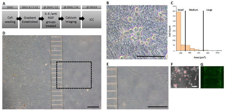
Isolation of axon-like structures in adult mouse DRG neuron cultures in microfluidic-based culture platforms (A) Experimental timeline. (B) Cell viability observations in phase contrast microscopy depicting healthy neuronal cell bodies. (C) Dorsal root ganglia cross-sectional area categorization with the majority of cells being of small-medium range. (D) Representative image of a seeded device with the soma (left) and axonal (right) chambers interconnected via microchannels at DIV 6 (E) Magnified view of the dashed rectangle from panel D demonstrating isolated axon-like structures crossing microchannels. (F) Axon-like structures crossing identified by the fluorescent tracer, Dil, applied to the axonal chamber. Dil is taken up by crossing axons and retrogradely transported to the soma. (G) Axon-like structures stained with peripherin, a marker that is preferentially expressed by peripheral sensory neurons. Horizontal scale bars represent 150 μm.

