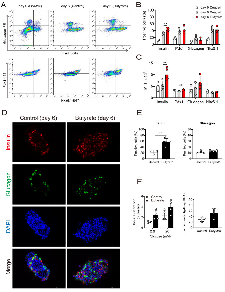
Figure 2.
Butyrate increased the number of insulin positive cells and insulin content. (A) FACS analysis of NPICCs before and after treatment with butyrate (1000 µM) for six days. NPICCs were dispersed into single cells and analyzed by intracytoplasmic staining for insulin (AF647), glucagon (PE), Pdx1 (AF488) and Nkx6.1 (AF647). (B,C) Butyrate increased the number of insulin positive cells and the median fluorescence intensity (MFI) of insulin stained cells. (D,E) Immunofluorescence staining for insulin (red) and glucagon (green) confirmed increase of insulin positive NPICCs in the butyrate treated group (1000 µM for six days). scale bars = 100 μm. (F) GSIS and Insulin content were moderately increased in NPICCs treated with butyrate. Data are presented as mean ± SD (B,E,F) or median ± SD (C) of three to six independent experiments. ** p < 0.01 vs. control group.

