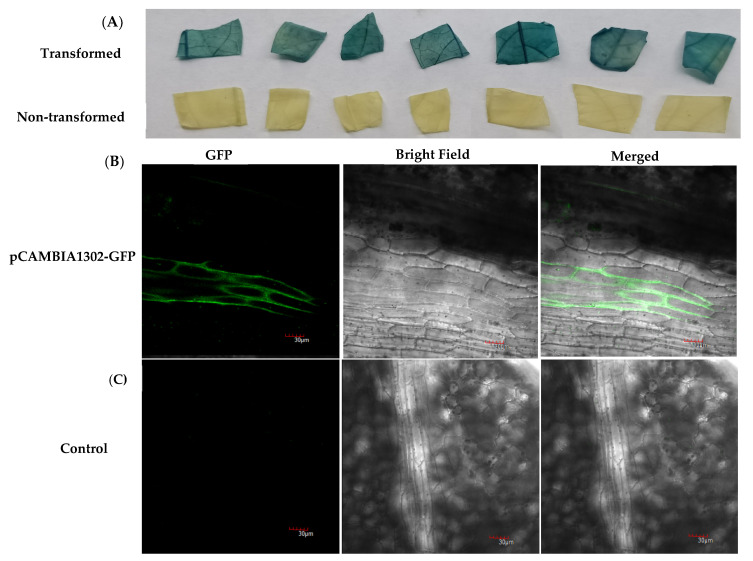
Figure 3.
Histochemical GUS staining and visualization of GFP fluorescence in transgenic plants. (A) Histochemical β-glucuronidase (GUS) staining of leaf segments of different transgenic plants and non-transgenic control plants. (B,C) Visualization of GFP reporter gene in transgenic (pCAMBIA1302) and non-transgenic (control) passion fruit plant leaves under laser scanning confocal microscope. (B) GFP expression on transformed passion fruit plant leaf; (C) non-transformed plant leaf as control; all bars are 30 µm.

