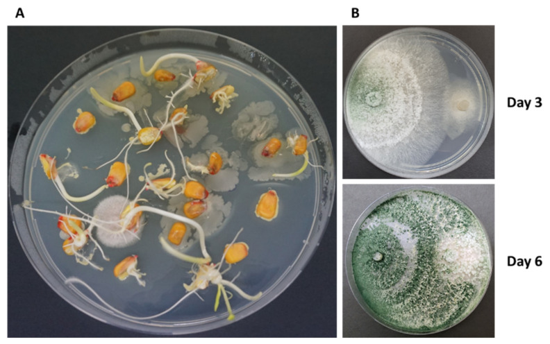
Figure 8.
Maize seeds microflora. (A) Maize grains were disinfected externally, cut lengthwise, and placed on a potato dextrose agar (PDA) medium with the cutting surface turned downwards. Petri dishes were maintained in the dark at 28 ± 1 °C for 2–3 days. (B) Plate mycoparasitism assays—T. Asperellum (P1, left side) vs. Magnaporthiopsis maydis (right side) [69].

