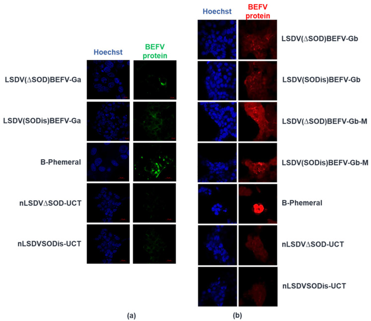
Figure 4.
BEFV protein expression detected by immunofluorescence. MDBK cells grown on chamber slides were infected with B-Phemeral (positive control) and each of the vaccines at an MOI of 0.1 for 48 h. The two parent LSDV vaccines (nLSDV∆SOD-UCT and nLSDVSODis-UCT) were used as negative controls. Anti-B-Phemeral rabbit serum was used as the primary antibody for all samples (1:1000 dilution). (a) detection of BEFV Ga expression (green) using donkey anti-rabbit Alexa488 secondary antibody (1:500); (b) detection of BEFV Gb (red) and BEFV-Gb-M (red) using anti-rabbit CY3 secondary antibody (1:500). Nucleic acid was stained with Hoechst (blue).

