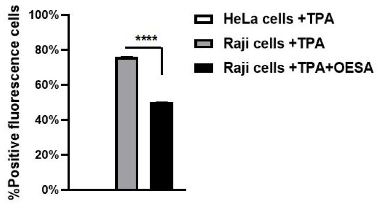
Figure 6.
Antiviral effect of OESA in the Raji cells line. Raji cells were stimulated with TPA, exposed to OESA (0.31 mg/mL), incubated with primary antibody, and labeled secondary antibody. The values represent the percentage of fluorescence cells. Asterisks indicate significant differences compared to OESA-untreated sample. (**** p < 0.0001). Results were expressed as mean ± standard deviations (n = 3).

