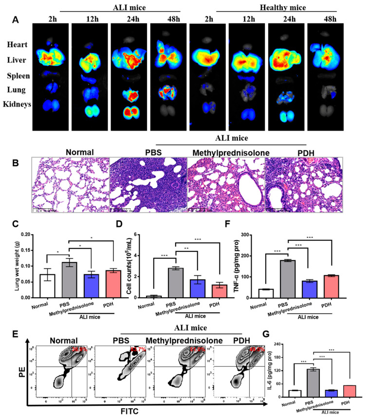
Figure 4.
In vivo therapeutic effect of PDH nanoparticles on ALI-induced mice. (A) Biodistribution of PDH/ICG in vivo. The fluorescence images of harvested organs from mice treated with LPS or not at 2 h, 12 h, 24 h and 48 h. (B) Morphologic alterations in lungs after different treatments were identified by hematoxylin and eosin staining. Scale bar = 200 µm. (C) The lung wet weight. (D) The total cell counts in the bronchoalveolar lavage fluid detected by flow cytometry. (E) The neutrophil counts in the bronchoalveolar lavage fluid measured by flow cytometry. (F) The level of TNF-α in lung tissues detected by TNF-α ELISA kit. (G) The level of IL-6 in lung tissues detected by IL-6 ELISA kit. (* p < 0.05, ** p < 0.01, *** p < 0.001, n = 3).

