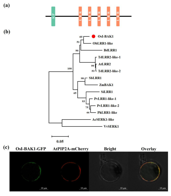
Figure 1.
Structure, phylogenetic relationship and subcellular localization of OsI-BAK1. (a) Schematic structure of OsI-BAK1. SP, signal peptide; LRR, leucin-rich repeat. (b) Sequence alignment of OsI-BAK1 and its homologs. Species abbreviations are included before the protein names: Ac, Ananas comosus; At, Aegilops tauschii subsp. strangulate; Bd, Brachypodium distachyon; Ob, Oryza brachyantha; Os, Oryza sative; Ph, Panicum hallii; Pv, Panicum virgatum; Sb, Sorghum bicolor; Si, Setaria italica; Td, Triticum dicoccoides; Vv, Vitis vinifera and Zm, Zea mays. Plant species and accession numbers from the NCBI database are as follows: AcSERK1-like, XP_020105022.1; AtLRR2, XP_020193068.1; BdLRR1, XP_003562364.1; ObLRR1-like, XP_006650233.1; OsI-BAK1, XP_015631230.1; PhLRR1-like, XP_025793013.1; PvLRR1-like-1, XP_039825633.1; PvLRR1-like-2, XP_039785598.1; SbLRR1, XP_002467662.1; SiLRR1, XP_004983941.1; TdLRR2-like-1, XP_037465236.1; TdLRR2-like-2, XP_037458478.1; VvSERK1, XP_002263235.1 and ZmBAK1, NP_001148919.1. The red dot indicates OsI-BAK1. The scale bar represents 0.05 amino acid substitution per site in the primary structure. (c) Subcellular localization of OsI-BAK1. Polyethylene glycol-mediated transient expression in rice protoplasts of OsI-BAK1-GFP and AtPIP2A-mCherry. OsI-BAK1-GFP, green fluorescent protein (GFP) fluorescence from OsI-BAK1-GFP; AtPIP2A-mCherry, mCherry fluorescence from AtPIP2A-mCherry; Bright, bright field and Overlay, co-localization of the OsI-BAK1-GFP and AtPIP2A-mCherry proteins. Bars, 10 μm.

